Endometriosis is a common condition that impacts on women’s health. Symptoms are variable and ultrasound and laparoscopy are the two most frequently used methods for the detection and management of endometriosis. In complicated cases MRI may complement ultrasound examination and assist in planning surgical management options.
Definition of Endometriosis
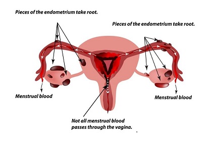
Endometriosis is defined as the presence of normal tissue of the lining of the uterine cavity (endometrium) in an abnormal place, usually the female pelvis. The most common sites in the pelvis are on and below the ovaries and deep in the pelvis behind the uterus, called the Pouch of Douglas. Here the endometriosis grows on the ligaments behind the uterus and on the vagina and rectum. It also may grow on the bladder, appendix, and even sometimes in the upper abdomen or in the abdominal wall scars of a laparoscopy or caesarean section.
Superficial deposits of endometriosis on the surface of the ovaries over time may lead to the development of ovarian endometriosis or endometriotic cysts. This will be recognisable on routine pelvic ultrasound.
Symptoms of Endometriosis
- Pain during periods (dysmenorrhoea)
- Pain with sexual intercourse (dyspareunia)
- Pain on defecation with periods (dyschezia)
- Chronic pelvic pain
- Abnormal bleeding
- Pain when urinating
- Infertility
- Pain with ovulation
- Fatigue
To make it confusing, some people with endometriosis have severe symptoms and others have very mild to sometimes hardly any symptoms. On the other hand, women who had the symptoms of endometriosis do not always have the disease.
Why does it occur?
The main mechanism believed responsible is backwards flow of menstrual blood through the Fallopian tubes into the pelvis during periods. The cells of the lining of the uterus (endometrial cells) can then implant in the pelvis and give rise to endometriosis lesions.
Next – Forms of Endometriosis, What Ultrasound Can and Cannot Diagnose

